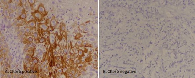
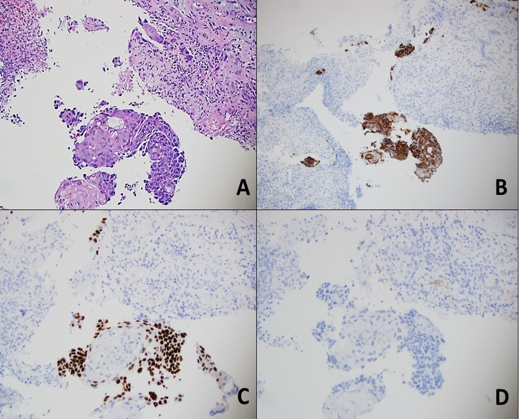
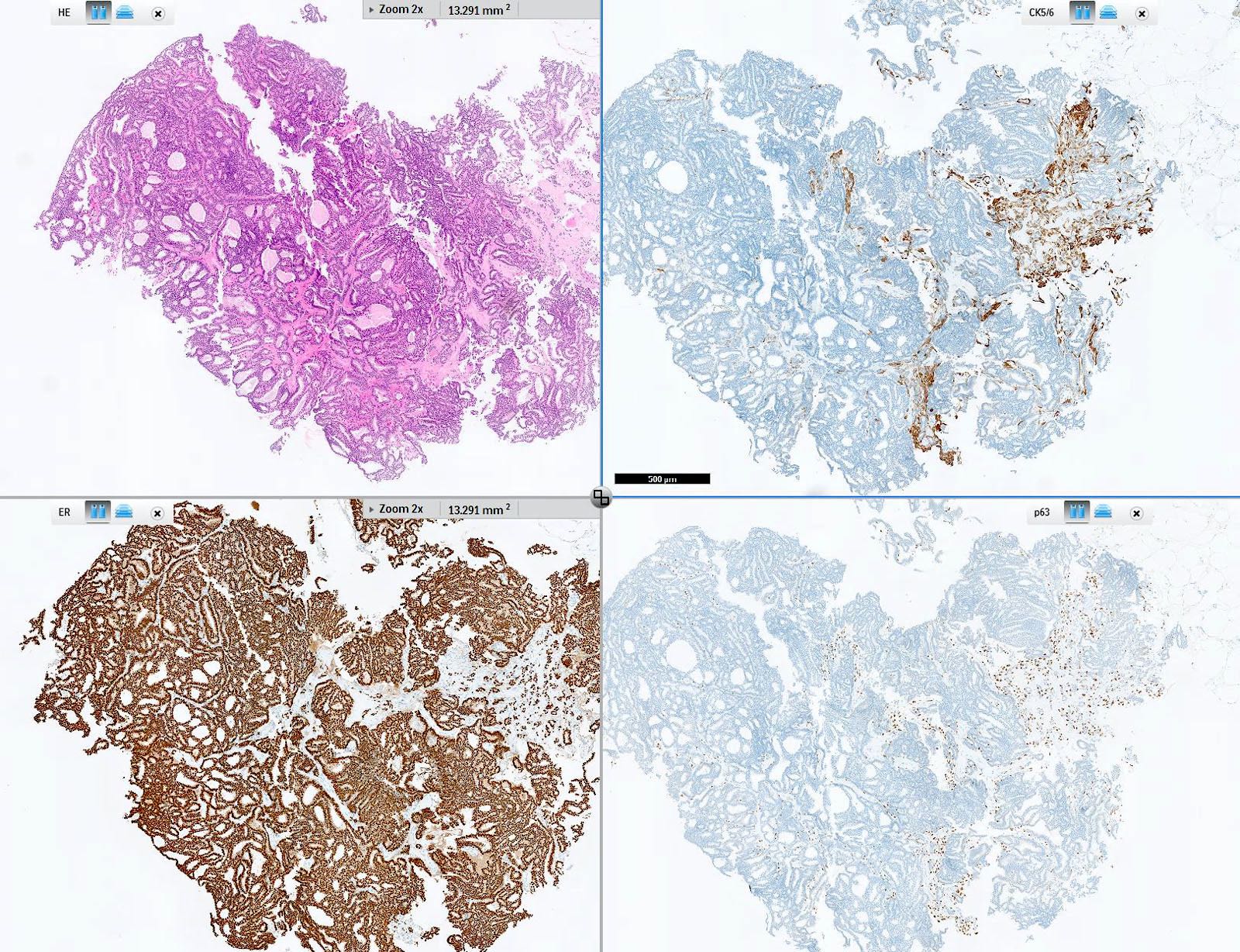
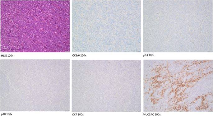
Tumors were scored as positive (>1% tumor cells reactive) or negative, and percentage of reactive tumor cells was recorded Results Napsin A, p63, p40, and CK5/6 were consistently negative in Different labs have different cutoff points for calling the cancer either "hormonereceptorpositive" or "hormonereceptornegative" For example, if less than 10% of your cells — or fewer than 1 in 10 — stain positive, one lab might call this a negative result Another lab might consider this positive, even though it is a low result Positive CK5/6 expression was noted in 63% (8 cases) and 134% (17 cases) revealed focal positive CK 5/6 expression On the other hand, 803% (102 cases) showed negative CK5/6 staining Significant association of CK5/6 expression was noted with tumor grade and muscularis propria invasion, however no significant association was noted with
Citoqueratinas 5 6 Ck 5 6 Histopat Laboratoris
Can cancer cause high ck levels
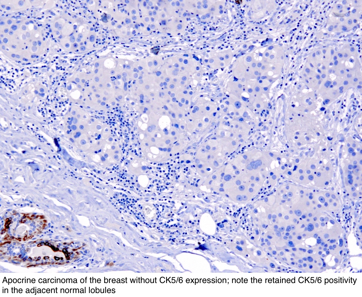
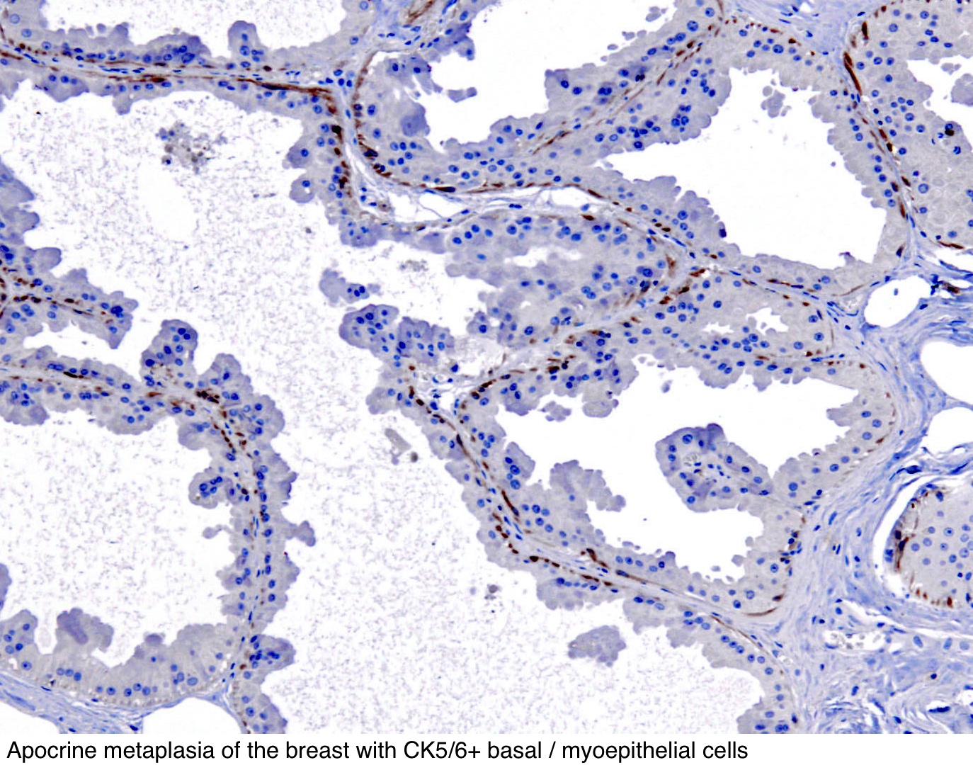
Can cancer cause high ck levels-These tests are sometimes used to help see if a tumor that includes the surface of the lung is a mesothelioma (see below) or an adenocarcinoma of the lungCK 5/6 expression breast carcinoma implies a 'basal like' molecular phenotype and is associated with poor prognosis This antibody is also used as a component of panels to differentiate benign and malignant breast lesions
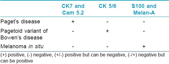


Indian Journal Of Dermatology Venereology And Leprology Practical Immunohistochemistry Of Epithelial Skin Tumor
All the 12 nonsmall cell lung cancers whose certain diagnosis was adenocarcinoma were positive for TTF1 TTF1, CK 5/6 and p63 seem to be useful forNonsquamous cell carcinomas of various primary sites, 141;Atins CK 5 and 6 and 34 βE12) in conjunction with estrogen receptor (ER) IHC may be helpful in these borderline cases Atypical proliferative lesions and in situ carcinoma lack staining with such keratins or they show occasional staining alone By contrast, a strong and diffuse staining pattern indicates a benign process (Fig 1A)4,5
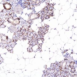
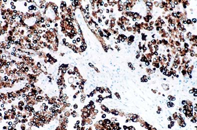
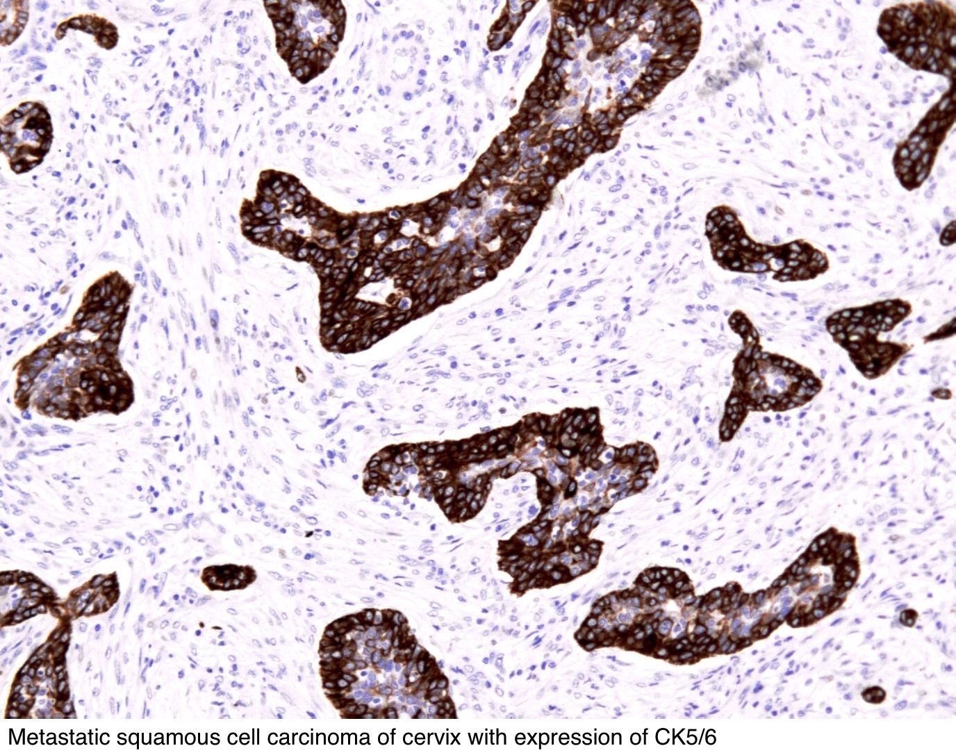
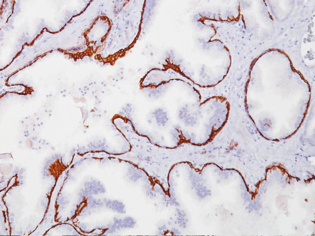
Immunostains for p63, CK 5/6, CK 7, and CK were performed and graded as follows 1, 50% of tumor cells stained Results Twenty of 22 PCAN expressed p63 and CK 5/6 Four of 15 and two of 15 MC were positive for CK 5/6 and p63, respectively Thirteen of 22 PCAN and 13 of 15 MC were positive for CK 7, respectively The p63 CK5 antibody multiplex stain has been especially designed for squamous cell carcinomas, particularly those derived in lung cancer Inhouse studies has shown greater than 80% of squamous cell carcinoma of the lung were positive and other studies have shown that the combination of p63 and CK5 was useful for differentiating adenocarcinoma from squamous cellBy Rodney T Miller, MD Cytokeratin 5/6 (CK 5/6) is a very useful antibody in diagnostic Surgical Pathology One of the most important aspects of this marker is that it characteristically stains squamous carcinomas strongly and diffusely, but generally stains adenocarcinomas focally, weakly, or not at all As such, it can be used as a marker of squamous differentiation
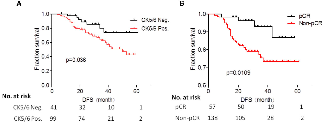
Fiftynine (656%) of 90 tumors were LCKpositive, and 19 (211%) were HCKpositive The incidence of LCK positivity was inversely correlated with nuclear or histological grade, however, theThese cells were strongly positive for CK7, GATA3, patchy positive for ER, and negative for CDX2, CK, TTF1, thyroglobulin, mammaglobin, Melanin A, and PAX8 IHC stains The interpretation based on these findings was metastatic breast adenocarcinoma The patient received tamoxifen for stage 4 ERpositive breast cancer, in spite of no clinical Nielson et al suggested that CK5/6 positive breast cancer have worse prognosis independent of tumor grade, Tstage and hormonal/Her2neu status 21 Similarly in another study it was proposed that CK5/6 is marker of shorter disease free survival, independent of other prognostic factors of breast cancer 8
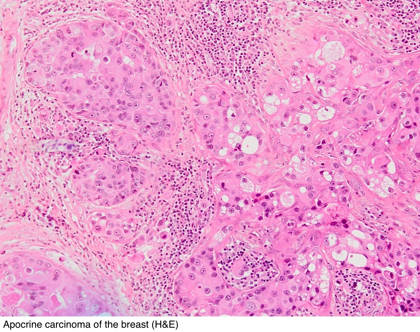


Pathology Outlines Cytokeratin 5 6 And Ck5


Academic Oup Com Ajcp Article Pdf 130 5 724 Ajcpath130 0724 Pdf
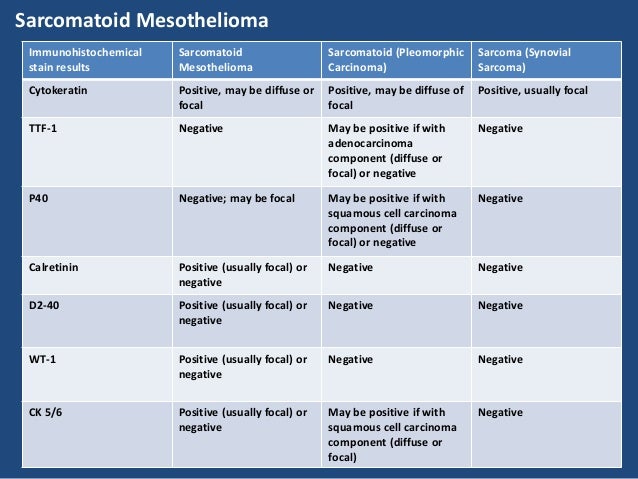
CK5/6 expression on the other hand is expressed in the basal cells in all lowgrade and some highgrade urothelial carcinomas Diffuse CK staining accompanied by loss of CK5/6positive basal layer is usually associated with aggressive clinical behavior Cytokeratin 5/6 is a positive marker for malignant pleural mesothelioma, found in more than threefourths of cases It is also found in certain types of lung cancers and breast cancers Pathologists use cytokeratin 5/6 to stain cancer tissue samplesThe diagnostic value of TTF1, CK 5/6, and p63 immunostaining in classification of lung carcinomas This study was aimed to evaluate the utility of a panel of antibodies, consisting of thyroid transcription factor1 (TTF1), p63, and cytokeratins (CK) 5/6 for distinguishing between small cell lung carcinoma (SCLC) and nonsmall cell lung carcinoma, as well as for identifying
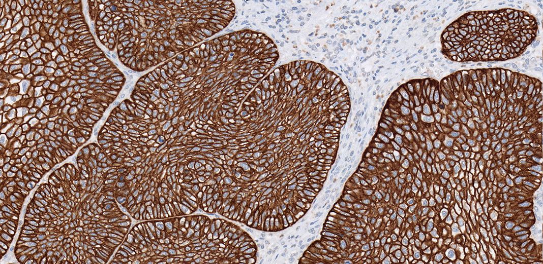


Lung Cancer Ihc Portfolio
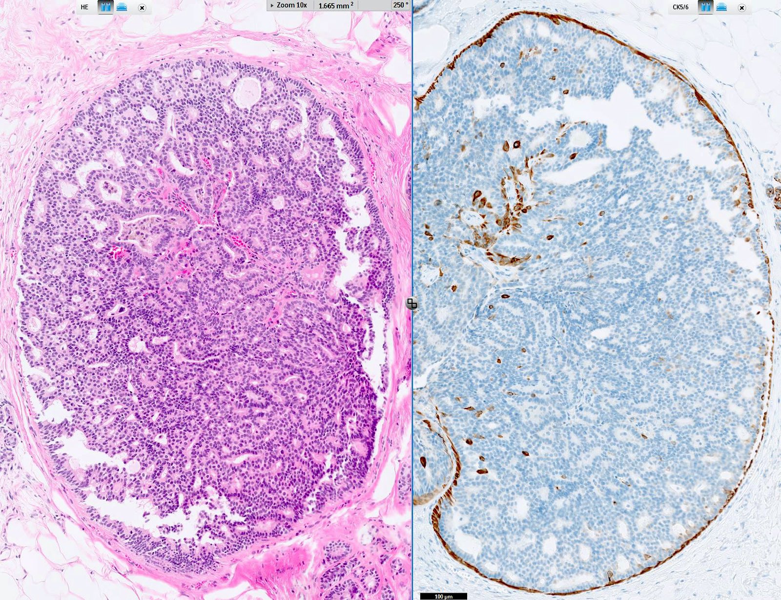


Pathology Outlines Cytokeratin 5 6 And Ck5
6%), showing different staining patterns from scattered stained cells to a diffuse staining, at times with a CK7 / CK in carcinomas of adrenal cortex and prostate ( Mod Pathol 00; ) To distinguish primary lung carcinoma (CK7 / CK) from metastatic colonic carcinoma to lung (CK7 / CK, BMC Cancer 06;631) To help distinguish colon carcinoma (80% are CK) and poorly differentiated prostatic carcinoma (90% are CK) at biopsySW480 cells were serially diluted 10fold into blood from healthy volunteers The final concentrations of SW480 cells were 10 6, 10 5, 10 4, 10 3, 10 2, 10 1, and 1/10 mL blood, respectively, and they were captured using the same refined protocol as above in triplicates for CK immunocytochemical stainingThe number of CKpositive SW480 cells for each


Lung Squamous Cell Carcinoma Associated With Hypoparathyroidism With S Ott


Cytokeratin 5 6 Antibody Ep24 Ep67 Bio Sb
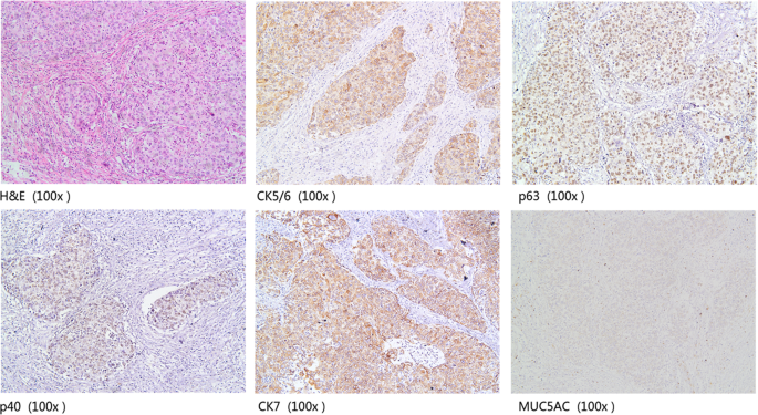
In the ER negative cases, 60% and 45% were positive for CK5/6 and CK14, respectively, whereas 393% and 303% of PR negative cases showed positivity for CK5/6 and CK14, respectively In contrast, only 303% of ER positive tumors were positive for CK5/6 and CK14 expression None of the cases which were PR positive showed expression of basal CKs For CK5/6, CK7, and MUC5AC, more than 5% of tumor cells with a membrane or cytoplasmic brownyellow granules were considered positive For p63 and p40, the positive standard was that more than 5% of tumor cells have brownyellow granules in the nucleus Statistical analysis Tumors with increased levels of HER2/neu are referred to as HER2positive The cells in HER2positive breast cancers have too many copies of the HER2/neu gene, resulting in greater than normal amounts of the HER2 protein These cancers tend to grow and spread more quickly than other breast cancers
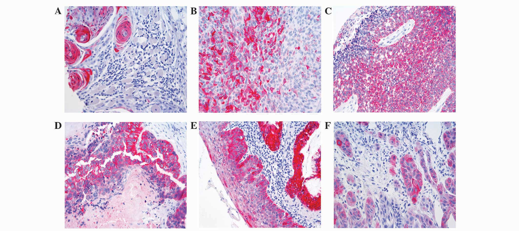


Cytokeratin And Protein Expression Patterns In Squamous Cell Carcinoma Of The Oral Cavity Provide Evidence For Two Distinct Pathogenetic Pathways
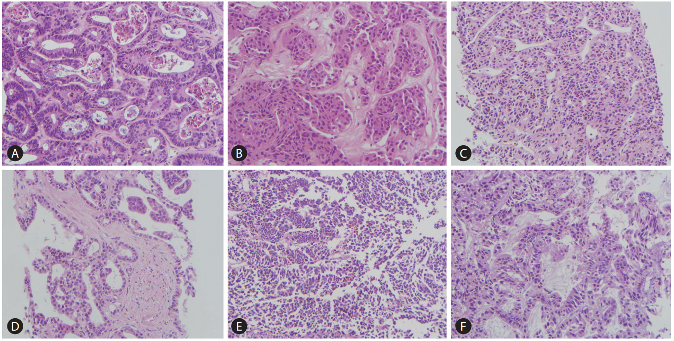


Pathologic Differential Diagnosis Of Metastatic Carcinoma In The Liver
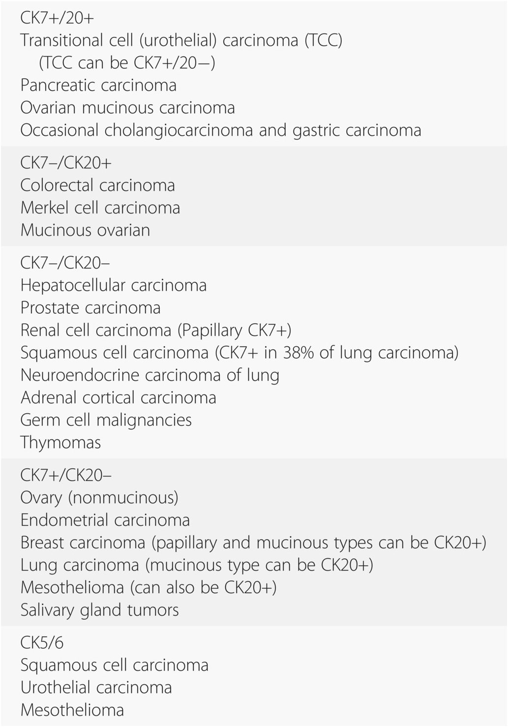
Most tumors demonstrated at least partly positive for cytokeratin 5/6 (n = 148; In breast cancer, expression of CK5/6 indicates a basal epithelial phenotype, 14 being especially frequent in BRCA1related breast cancer and associated with highgrade tumors and decreased survival 15 In endometrial carcinomas, expression of CK5/6 has not been evaluated in relation to clinicopathologic features or prognosis The aim of our study was to examine the associations between p63 or CK5/6 expression and tumor Therefore, ER cannot be a single mark to distinguish these tumors Secondly, adenocarcinomas of the breast, ovary and gastrointestinal tract show different CK7 and CK expression patterns In breast and ovary adenocarcinomas, CK7 is positive and CK is negative In contrast, CK is positive and CK7 is negative in gastrointestinal ones 5, 6
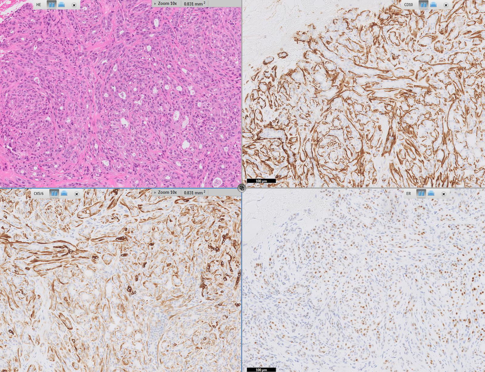


Pathology Outlines Cytokeratin 5 6 And Ck5
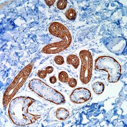


Cytokeratin 5 6 Ck 5 6 Pathology Resident Wiki Fandom
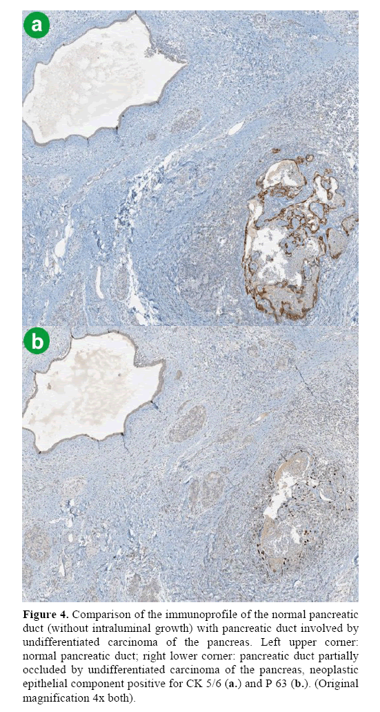
Abstract To facilitate the differential diagnosis of poorly differentiated metastatic carcinomas of unknown primary site, we evaluated p63 and cytokeratin (CK) 5/6 as immunohistochemical markers for squamous cell carcinomas The study cases were as follows squamous cell carcinoma of the lungs, head/neck, esophagus, cervix uteri, or anal canal, 73;It will say a tumor is hormone receptorpositive if at least 1% of the cells tested have estrogen and/or progesterone receptors Otherwise the test will say the tumor is hormone receptornegative Hormone receptorpositive (or hormonepositive) breast cancer cells have either estrogen (ER) or progesterone (PR) receptors or both These breast cancers can be treated with hormone therapyWatch the full video on IGTV Combination of CK7 and CK is vastly used to determine the primary site in a metastatic tumor of unknown originThe patte


Immunohistochemical Detection Of Cytokeratin 5 6 In Breast Cancer Download Scientific Diagram
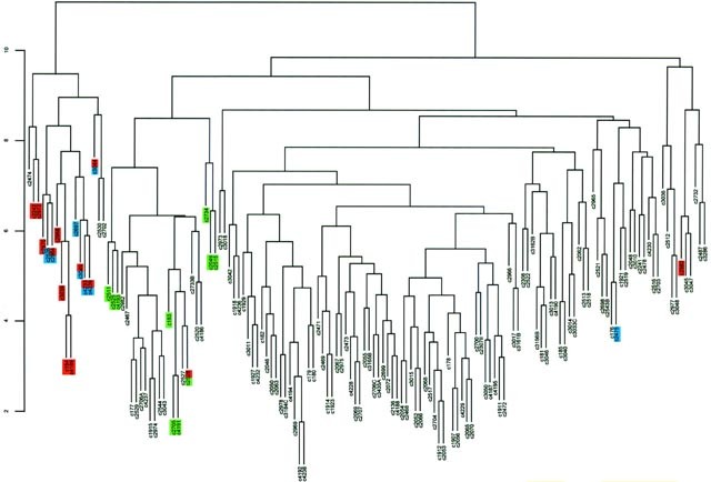


Cytogenetic Alterations And Cytokeratin Expression Patterns In Breast Cancer Integrating A New Model Of Breast Differentiation Into Cytogenetic Pathways Of Breast Carcinogenesis Laboratory Investigation
What does it mean if my report mentions special tests such as D240 (podoplanin), calretinin, WT1, BAP1, CEA, cytokeratin (CK) 5/6, HBME1, BerEP4, TTF1, and/or CD15 (LeuM1)?Primary lung squamous cell carcinomas expressed CK5, p40, and p63 in 94%97% of cases, whereas 44% were positive for CK7, % for GATA3, 7% for CDX2, and 3% for TTF1 Rare cases expressed PAX8, CK, or napsin A Pulmonary metastases of colorectal cancer were positive for CK in % and CDX2 in 99% of the cases With 6 cores per cancer case, a maximum of 72 cases were placed on each recipient TMA block Nonmalignant tissues (spleen, liver, kidney, and placenta) were included as controls for orientation at the top and bottom of each TMA block The entire 1995 Hawaii colorectal tissue collection was arrayed in a total of 5 TMA blocks
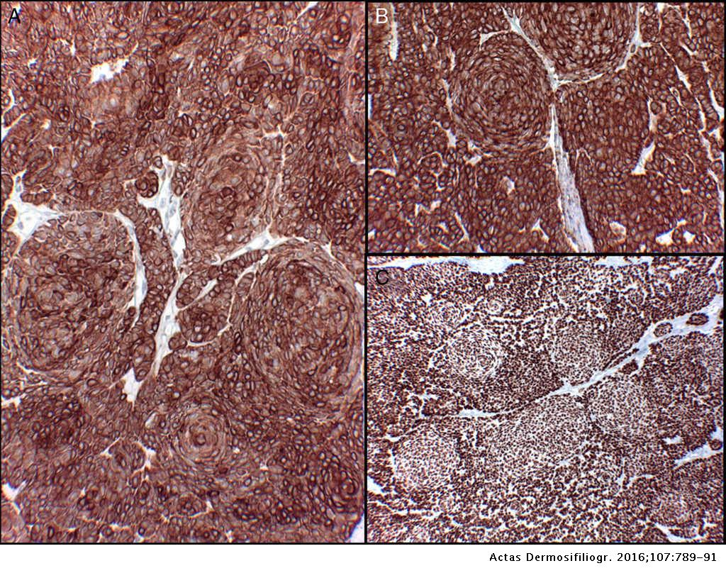


Trichogerminoma A Neoplasm With Follicular Differentiation And A Characteristic Morphology Actas Dermo Sifiliograficas


Mesothelioma Positive For Pan Ck Ck 5 6 Calretinin Wt1 Diagnostic Biosystems Immunohistochemistry Primary Antibodies Monoclonal Antibodies Polyclonal Antibodies Fitc Antibodies Automated Staining Instrumentation Mouse And Rabbit
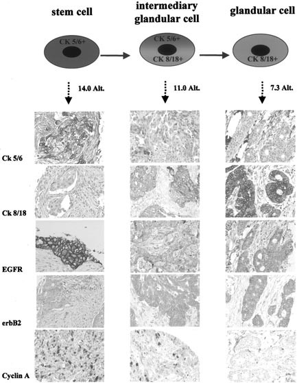
However, the need for a sensitive and specific panel of antibodies to differentiate lung adenocarcinoma from lung SqCC is of the utmost importance Thrombomodulin (CD141), p63, 34betaE12 and CK5/6 have been shown to be sensitive markers for SqCC Breast cancers are generally thought to express either luminal (CK8/18/19) or basal (CK5/14) cytokeratins 5, 9, 10 However, some CK5/14 and CK8/18coexpressing tumors have also been found 2 It is possible that certain papillary carcinomas derived from the papillomas might result from the CK 8/18positive tumor cells that replace the CK 5/6 positive cells The two models are complementary in explaining tumor heterogeneity and progression However, the hypothesis that the papillary carcinoma cells could be derived from the papilloma


Characterization Of Dll3 Positive Circulating Tumor Cells Ctcs In Patients With Small Cell Lung Cancer Sclc And Evaluation Of Their Clinical Relevance During Front Line Treatment Lung Cancer


The Use Of Immunohistochemistry Improves The Diagnosis Of Small Cell Lung Cancer And Its Differential Diagnosis An International Reproducibility Study In A Demanding Set Of Cases Sciencedirect
Coexpression of p63 and CK5/6 had a sensitivity of 077 and a specificity of 096 for squamous cell carcinomas Increasing the minimal criterion of positive immunostaining for both markers to more than 50% of immunoreactive tumor cells resulted in a specificity of 099, although the sensitivity diminished to 066Cytokeratin (CK) and cytokeratin 7 (CK7) immunohistochemistry was used to validate the utility of the TMA, and the expression of these proteins was correlated with patient and tumor characteristics The TMA comprised tissue specimens from 286 patients representing 47% of all invasive cases diagnosed statewide in 1995 A cohort of 215 patients with highgrade serous ovarian carcinoma was evaluated immunohistochemically for the protein expression of cytokeratin 5/6 Most tumors demonstrated at least partly positive for cytokeratin 5/6 (n = 148;


Pathology Outlines Cytokeratin 5 6 And Ck5
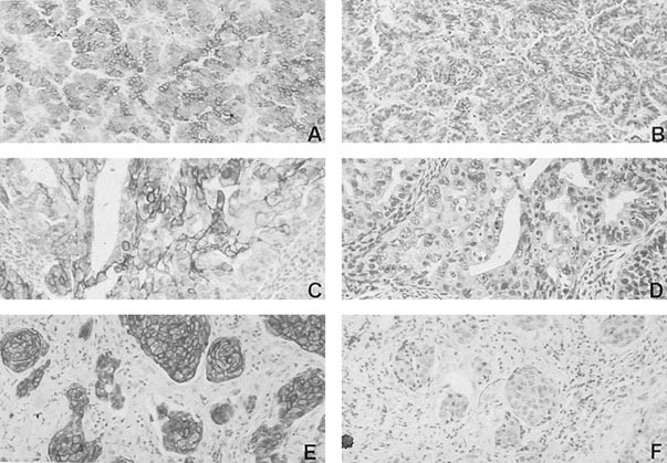


Expression Of Cytokeratin 5 6 In Epithelial Neoplasms An Immunohistochemical Study Of 509 Cases Modern Pathology
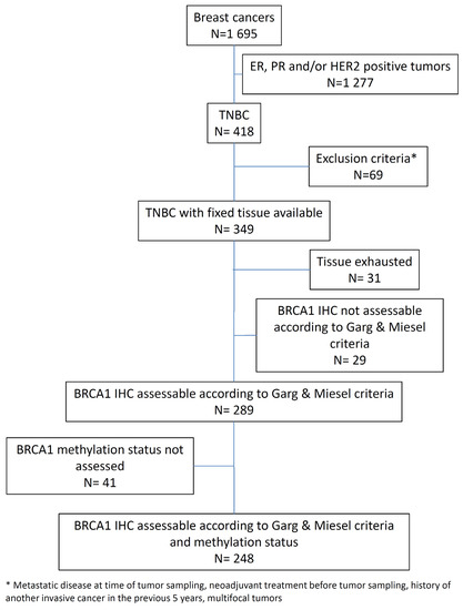
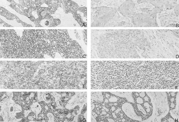
6%), showing different staining patterns from scattered stained cells to a diffuse staining, at times with a distinctive tumorstroma border motif Sixtyseven (31%) were entirely negativeCK 5/6 was expressed in 7777% of SCCs and CK 7 was expressed in 9259% of ACs Synaptophysin and chromograninA were expressed in 100% of neuroendocrine (NE) carcinomas The tumor cells were CK5/6 positivity helps define a basallike subtype of triple negative breast carcinoma (TNBC) with poorer prognosis (Clin Cancer Res 06;);


Biological Significance Of Gata3 Cytokeratin Cytokeratin 5 6 And P53 Expression In Muscle Invasive Bladder Cancer


Cytokeratin Wikipedia
Cyclin D1 and CK5/6 staining could be used in concert to distinguish between the diagnosis of papilloma (Cyclin D1 3700%, CK 5/6 negative) In the lung, distinguishing epithelioid mesothelioma (CK5/6 positive in %) from lung adenocarcinoma (CK5/6 negative in 85%) An adenocarcinoma showing typical presentation of the breast profile by immunohistochemistry Hematoxylineosin stain (left upper), cytokeratin (CK)7 (right upper), CK (left lower), and mammaglobin (right lower) CK7 and mammaglobin are positive in the cytoplasm of tumor cells (original magnification ×0) Figure 3 CK5/6 and CK5/6 cases did not significantly differ with respect to patient age, frequency of PR expression, tumor size, rate of axillary node involvement, or HER2/neu overexpression In summary, the present study demonstrated that 17% of ILCs express CK5/6, and that CK5/6 cases are more likely to be ER and have a high modified


Ispub Com Ijpa 13 2


Cutaneous Epithelial Tumors Dog Figure 5 Basal Cell Carcinoma Tumor Download Scientific Diagram
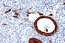
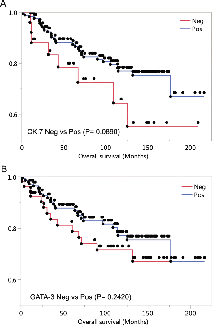
Cytokeratins are a family of intermediate filaments that are predominantly found in cells of epithelial origin Keratins can be divided into two types acidic cytokeratins (Type I) and basic cytokeratins (Type II)1 1 Practical Uses 2 Antibodies 21 AE1/A 22 CAM 52 23 CK5/6 24 CK7 25 Cytokeratin 14 (CK 14) 26 Cytokeratin 17 (CK 17) 27 Cytokeratin 18 (CK 18) 28 Cytokeratin No dermal invasion is identified Immunohistochemical studies show the cells to be positive for CK7, CAM52 and HER2 while negative for SOX10, p63, CK5/6, CK and adipophilin The morphologic and immunophenotypic findings are Among Grade 3 tumors, 17% of ER positive and 21% PR positive tumors were seen in CK7 negative/GATA3 positive tumors, in contrast to 1% of ER positive and 45% PR positive tumors being CK7 positive/GATA3 negative (P < ) (Table 4) Her2 expression did not have any association with CK7/GATA3 status (P = ) (Table 4)


Nuevos Aspectos Sobre La Iniciacin Y Progresin Del


Immunohistochemistry Stains In Squamous Cell Carcinoma And Download Scientific Diagram
High Ki67 indices were associated with negative CK (p = 0002) and diffuse CK5/6 (p = 0001) staining By contrast, tumors with diffuse GATA3 expression had Other authors adopted these criteria as a basis for defining basallike tumors Livasy et al reported that the most consistent profile of BLBC is ER–, HER2– and positive for EGFR, CK 5/6, CK 8/18 Basallike subtype represents 15–% of all breast cancers regardless of the method of analysis used in the study and ethnic groupThe diagnosis of prostate adenocarcinoma is aided by IHC staining for basal cell layer markers, such as p63, cytokeratin 5/6 (CK 5/6), and high molecular weight cytokeratin (CK HMW) as well as prostate'specific' markers This document will discuss the potentials and pitfalls of the individual markers used in the diagnosis of prostate cancer


07 Histology Cytology And Biomarkers In Pleural Diseases


Www Ajol Info Index Php Mjz Article View
Associated with premenopausal African American women (JAMA 06;), BRCA1 mutations (Mod Pathol 05;, J Natl Cancer Inst 03;9514) and higher propensity for brain metastases (AmTTF1, CK7 and p63 expression have been used to differentiate primary lung cancers;


2


Immunohistochemical And Clinical Characterization Of The Basal Like Subtype Of Invasive Breast Carcinoma Clinical Cancer Research


Cytokeratin 5 6 Ck 5 6 Pathology Resident Wiki Fandom


Cytokeratin 5 6 Antibody D5 16b4 Bio Sb


Immunohistochemistry In Prostate Pathology Pdf Free Download


Cytokeratin 7 Negative And Gata Binding Protein 3 Negative Breast Cancers Clinicopathological Features And Prognostic Significance Springerlink


Triple Negative Breast Carcinoma A Comparative Study Between Breast Lesion And Lymph Node Metastases A Preliminary Study


Scielo Brasil Lung Cancer Biopsy Can Diagnosis Be Changed After Immunohistochemistry When The H Amp E Based Morphology Corresponds To A Specific Tumor Subtype Lung Cancer Biopsy Can Diagnosis Be Changed After Immunohistochemistry


Cancers Free Full Text Brca1 Promoter Hypermethylation Is Associated With Good Prognosis And Chemosensitivity In Triple Negative Breast Cancer Html


Cytokeratin 5 6 Expression In Bladder Cancer Association With Clinicopathologic Parameters And Prognosis Bmc Research Notes Full Text


Cytokeratin 5 6 Expression In Benign And Malignant Breast Lesions Bhalla A Manjari M Kahlon S K Kumar P Kalra N Indian J Pathol Microbiol


Pathology Outlines Cytokeratin 5 6 And Ck5


Differential Diagnosis Of Metastatic Tumors Chapter 6 Silverberg S Principles And Practice Of Surgical Pathology And Cytopathology


Utility Of 10 Immunohistochemical Markers Including Novel Markers Desmocollin 3 Glypican 3 S100a2 S100a7 And Sox 2 For Differential Diagnosis Of Squamous Cell Carcinoma From Adenocarcinoma Of The Lung Journal Of Thoracic Oncology


1


Ispub Com Ijpa 13 2


Frontiers Analysis Of Ck5 6 And Egfr And Its Effect On Prognosis Of Triple Negative Breast Cancer Oncology


1


Immunohistochemistry Cytokeratin Ae1 Ae3 Strongly Positive Diffuse Download Scientific Diagram


Pathology Outlines Cytokeratin 5 6 And Ck5


Cureus Primary Gastric Squamous Cell Carcinoma


Ispub Com Ijpa 13 2


A Combination Of Cytokeratin 5 6 P63 P40 And Muc5ac Are Useful For Distinguishing Squamous Cell Carcinoma From Adenocarcinoma Of The Cervix Diagnostic Pathology Full Text


Pdf Expression Of Cytokeratin 5 6 In Epithelial Neoplasms An Immunohistochemical Study Of 509 Cases Semantic Scholar
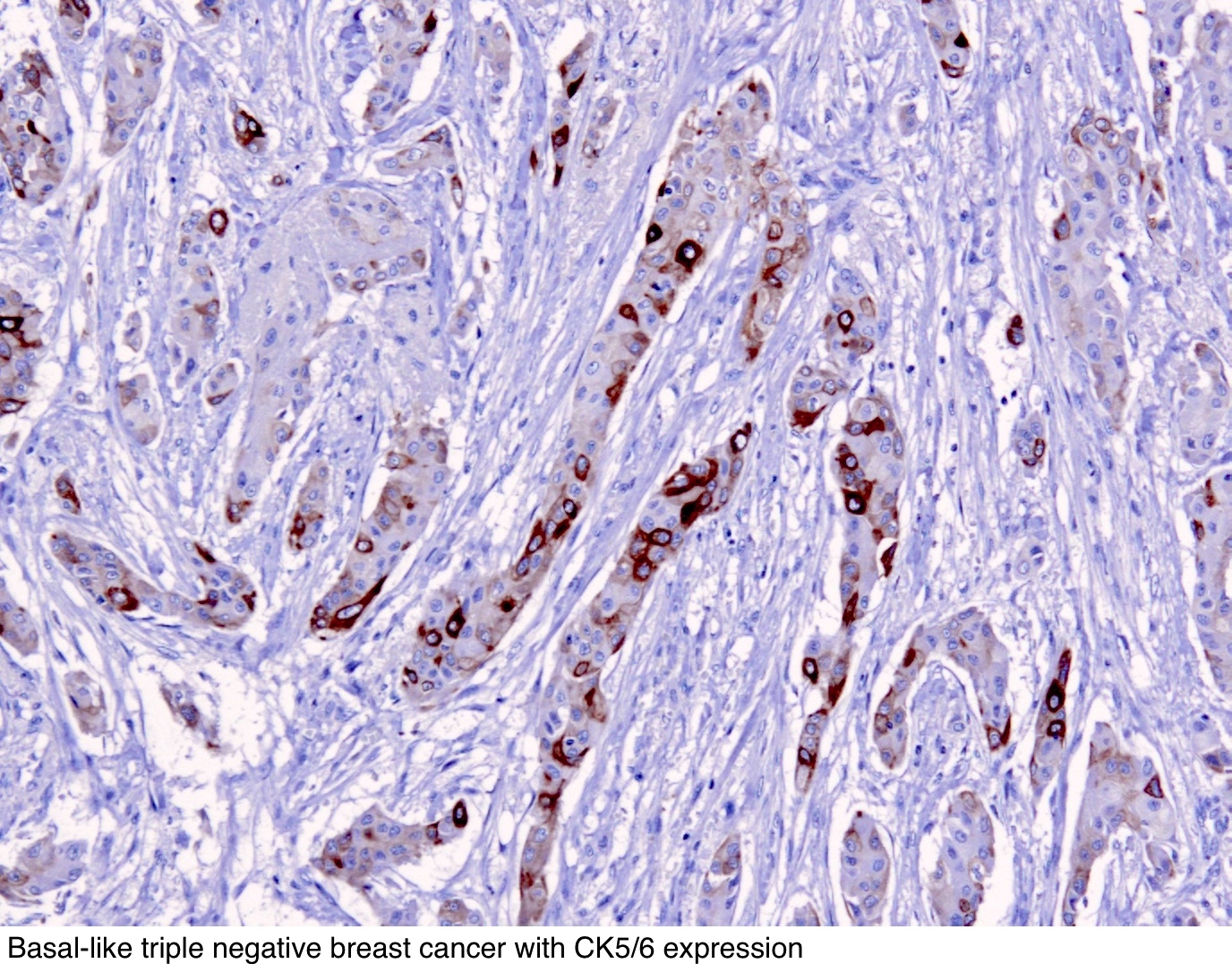


Pathology Outlines Cytokeratin 5 6 And Ck5


Correlation Of Epithelial Mesenchymal Transition And Ck19 Expression In Hepatocellular Carcinoma Kirimlioglu Journal Of Gastroenterology And Hepatology Research


Expression Of P63 And Ck5 6 In Early Stage Lung Squamous Cell Carcinoma Is Not Only An Early Diagnostic Indicator But Also Correlates With A Good Prognosis Ma 15 Thoracic Cancer


Example Of A Basal Like Breast Cancer Stained With Cytokeratin 5 6 Download Scientific Diagram


Cytokeratin 5 Determines Maturation Of The Mammary Myoepithelium Sciencedirect
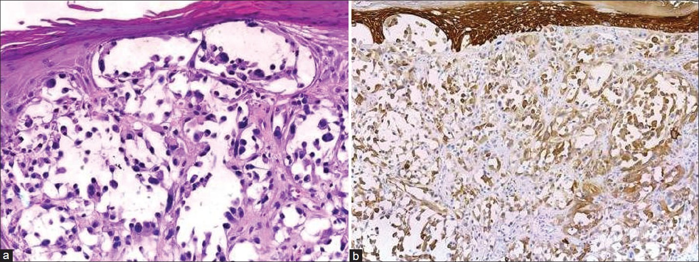


Indian Journal Of Dermatology Venereology And Leprology Practical Immunohistochemistry Of Epithelial Skin Tumor


Cytokeratin 5 6 Expression In Benign And Malignant Breast Lesions Bhalla A Manjari M Kahlon S K Kumar P Kalra N Indian J Pathol Microbiol


Indian Journal Of Dermatology Venereology And Leprology Practical Immunohistochemistry Of Epithelial Skin Tumor


Www Patologi Com Dako immun Prostate Pathology Pdf


Citoqueratinas 5 6 Ck 5 6 Histopat Laboratoris


Expression Of P63 And Ck5 6 In Early Stage Lung Squamous Cell Carcinoma Is Not Only An Early Diagnostic Indicator But Also Correlates With A Good Prognosis Ma 15 Thoracic Cancer


Mesothelioma Positive For Pan Ck Ck 5 6 Calretinin Wt1 Diagnostic Biosystems Immunohistochemistry Primary Antibodies Monoclonal Antibodies Polyclonal Antibodies Fitc Antibodies Automated Staining Instrumentation Mouse And Rabbit


Immunohistochemical And Clinical Characterization Of The Basal Like Subtype Of Invasive Breast Carcinoma Clinical Cancer Research
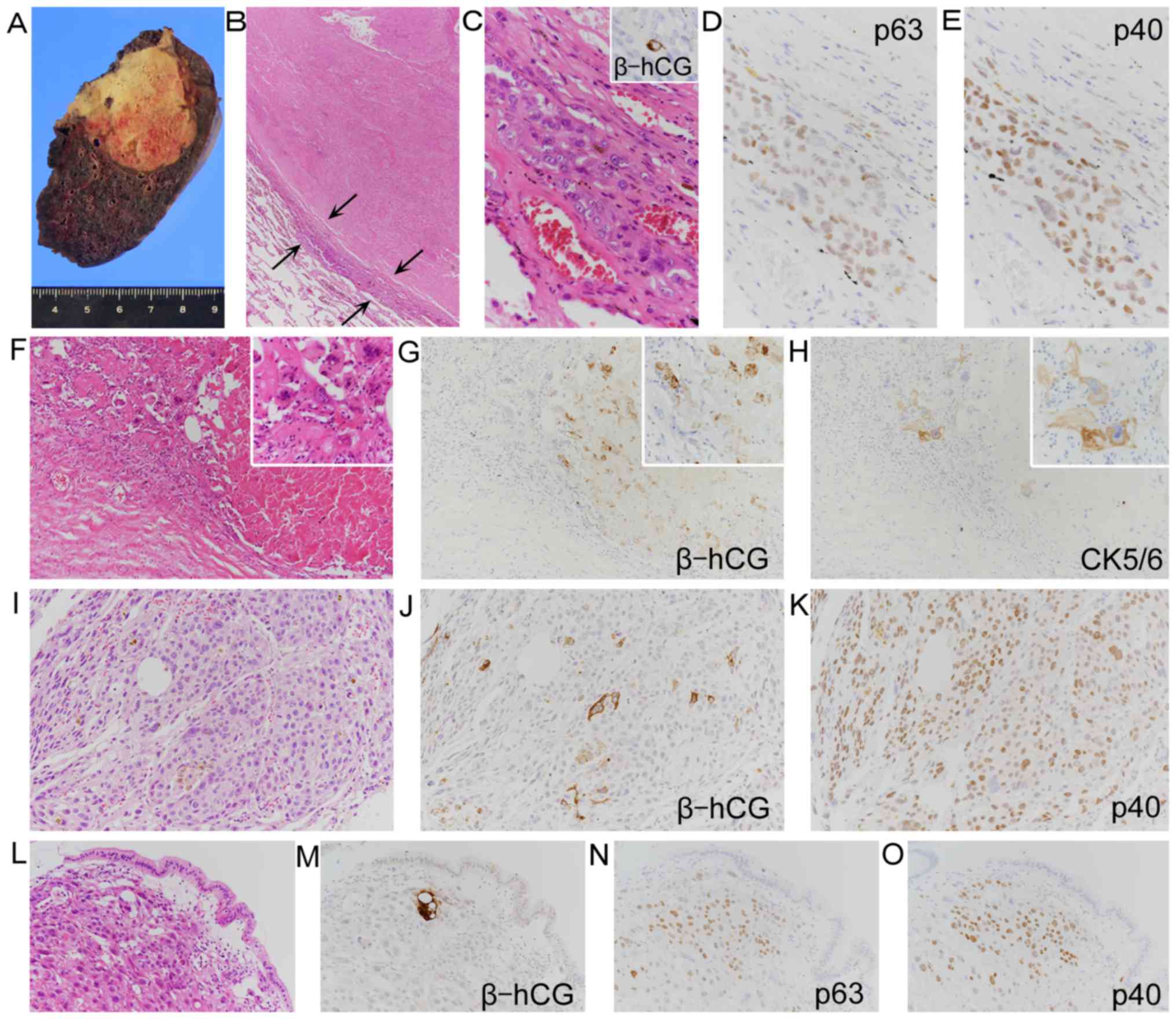


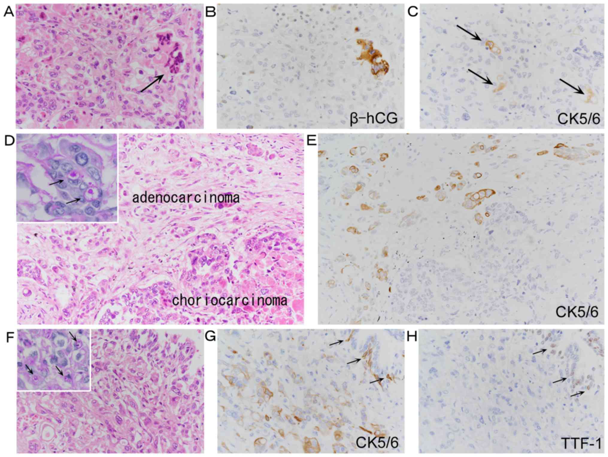
Focal Positivity Of Immunohistochemical Markers For Pulmonary Squamous Cell Carcinoma In Primary Pulmonary Choriocarcinoma A Histopathological Study


2
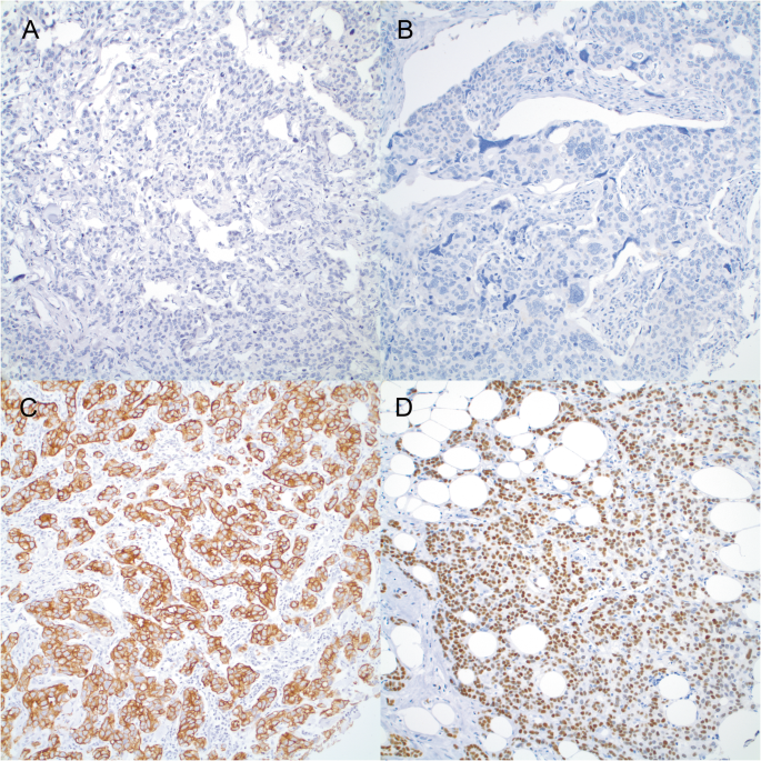


Cytokeratin 7 Negative And Gata Binding Protein 3 Negative Breast Cancers Clinicopathological Features And Prognostic Significance Springerlink


Pathology Outlines Cytokeratin 5 6 And Ck5


Cytokeratin 5 6 Antibody Biocare Medical


Triple Negative Breast Cancer Assessing The Role Of Immunohistochemic tt


Undifferentiated Anaplastic Carcinoma Of The Pancreas With Osteoclast Like Giant Cells Showing Various Degree Of Pancreas Duct Involvement A Case Report And Literature Review Insight Medical Publishing


A Combination Of Cytokeratin 5 6 P63 P40 And Muc5ac Are Useful For Distinguishing Squamous Cell Carcinoma From Adenocarcinoma Of The Cervix Diagnostic Pathology Full Text
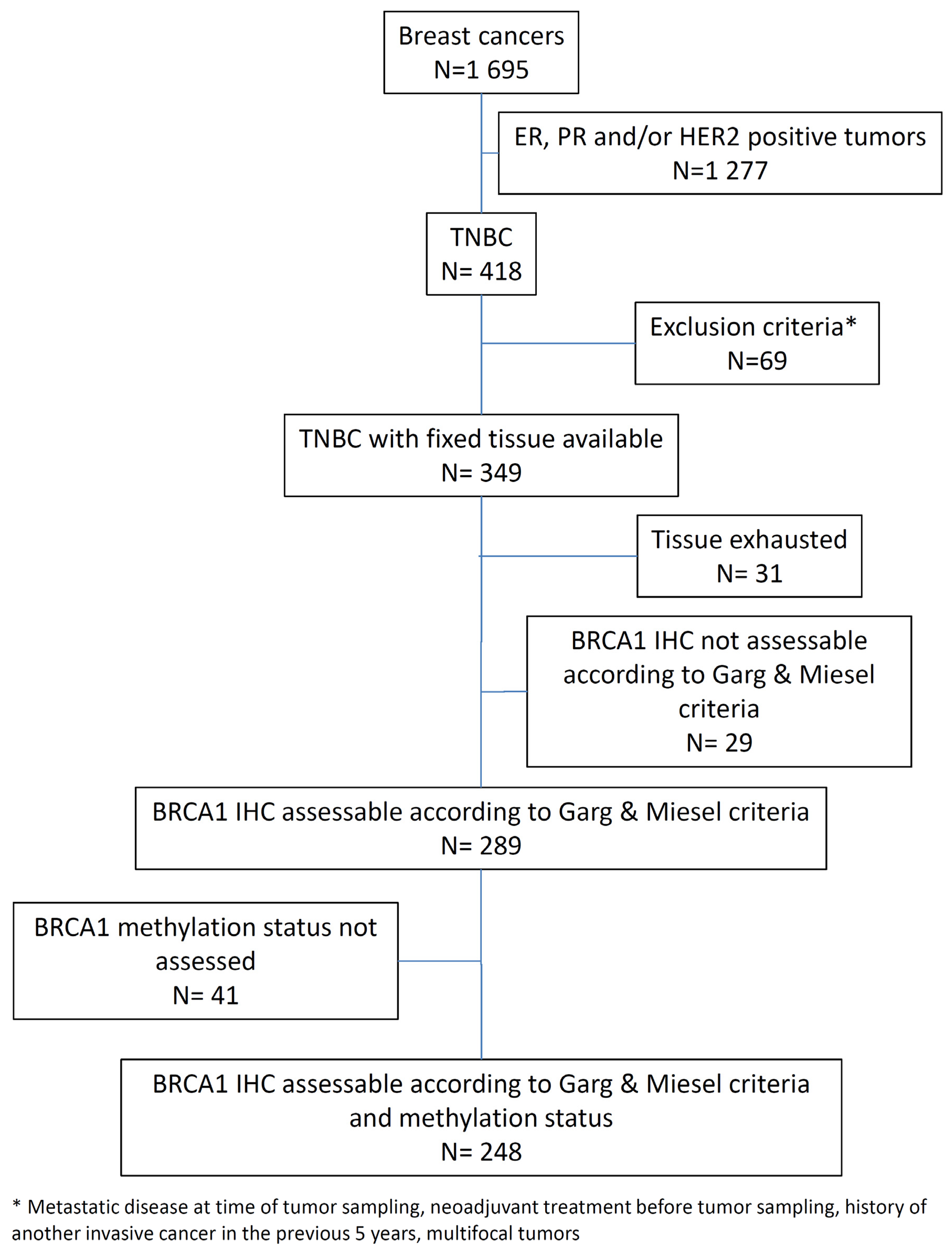


Cancers Free Full Text Brca1 Promoter Hypermethylation Is Associated With Good Prognosis And Chemosensitivity In Triple Negative Breast Cancer Html
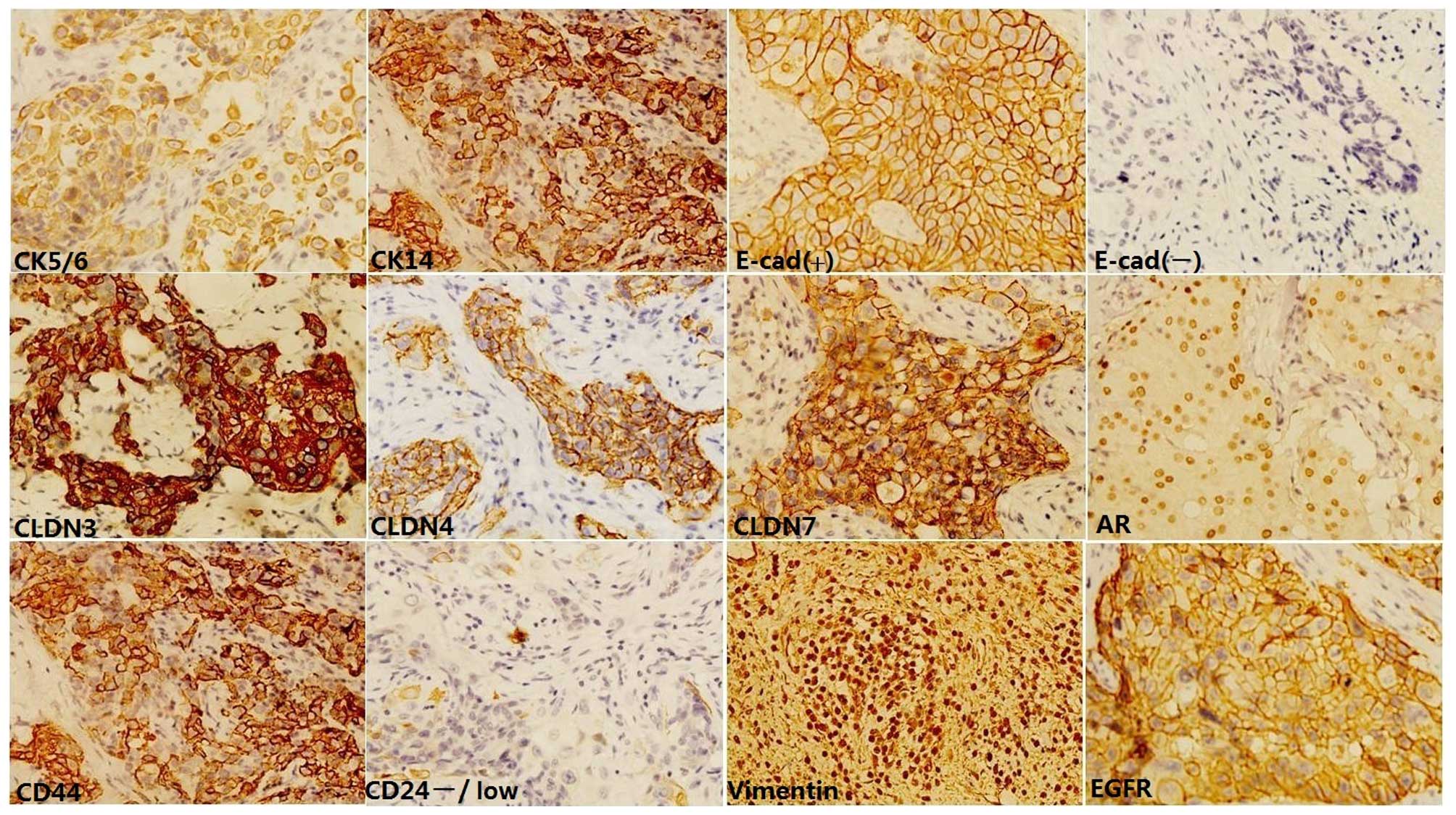


Attempt Towards A Novel Classification Of Triple Negative Breast Cancer Using Immunohistochemical Markers


Mesothelioma Positive For Pan Ck Ck 5 6 Calretinin Wt1 Diagnostic Biosystems Immunohistochemistry Primary Antibodies Monoclonal Antibodies Polyclonal Antibodies Fitc Antibodies Automated Staining Instrumentation Mouse And Rabbit


Hematoxylin Eosin He Staining And Immunohistochemistry The Tumor Download Scientific Diagram


Identification Of Brca1 Deficiency Using Multi Analyte Estimation Of Brca1 And Its Repressors In Ffpe Tumor Samples From Patients With Triple Negative Breast Cancer


Q Tbn And9gctuaeubeofickfpn5 Lfmiu2rtllphz Iclkv2zftgxliqt Avr Usqp Cau
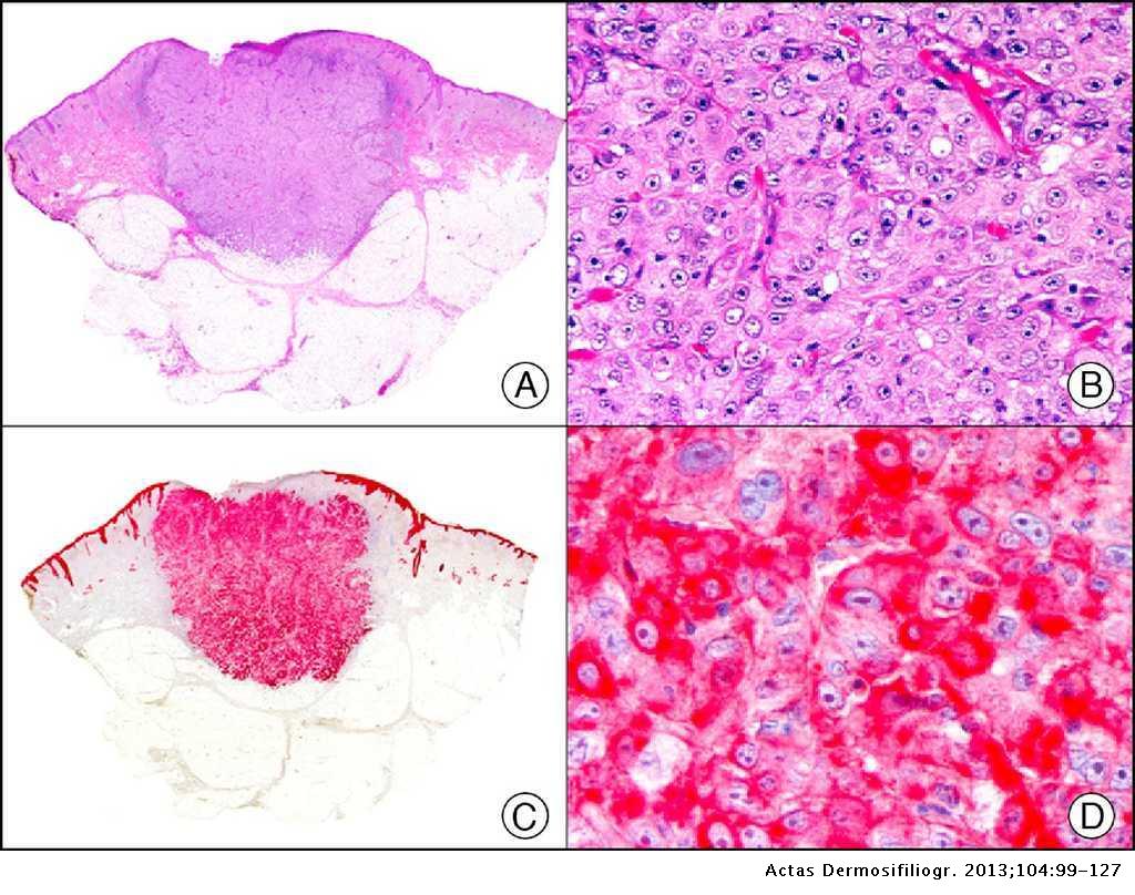


Immunohistochemistry In Dermatopathology A Review Of The Most Commonly Used Antibodies Part I Actas Dermo Sifiliograficas


1
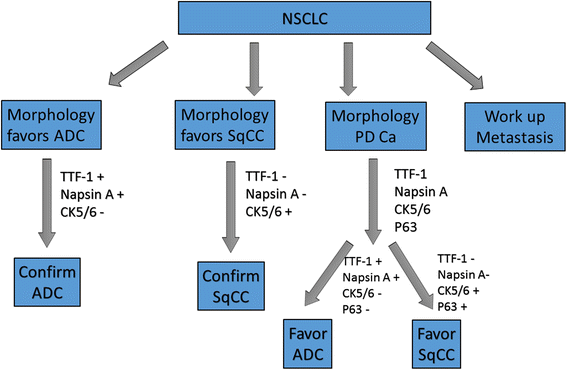


Utility Of Five Commonly Used Immunohistochemical Markers Ttf 1 Napsin A Ck7 Ck5 6 And P63 In Primary And Metastatic Adenocarcinoma And Squamous Cell Carcinoma Of The Lung A Retrospective Study Of 246 Fine


Scielo Brasil Comparison Of Nuclear Grade And Immunohistochemical Features In Situ And Invasive Components Of Ductal Carcinoma Of Breast Comparison Of Nuclear Grade And Immunohistochemical Features In Situ And Invasive


Immunohistochemistry In Dermatopathology A Review Of The Most Commonly Used Antibodies Part I Actas Dermo Sifiliograficas


D Nb Info 34
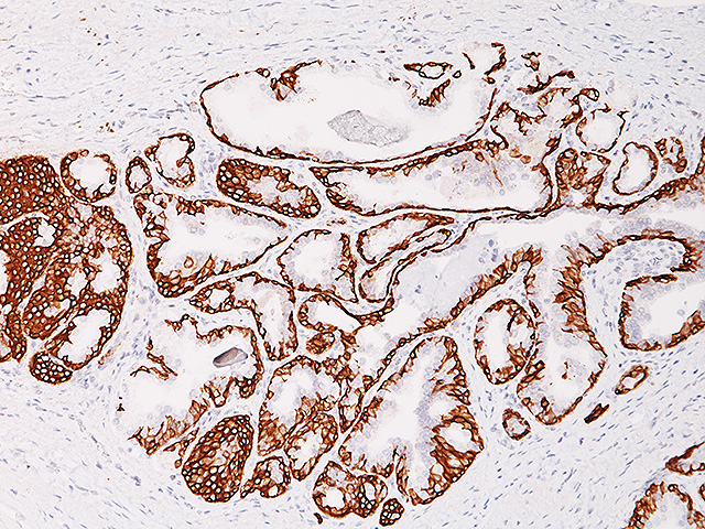


Cytokeratin 5 14 Antibody Biocare Medical
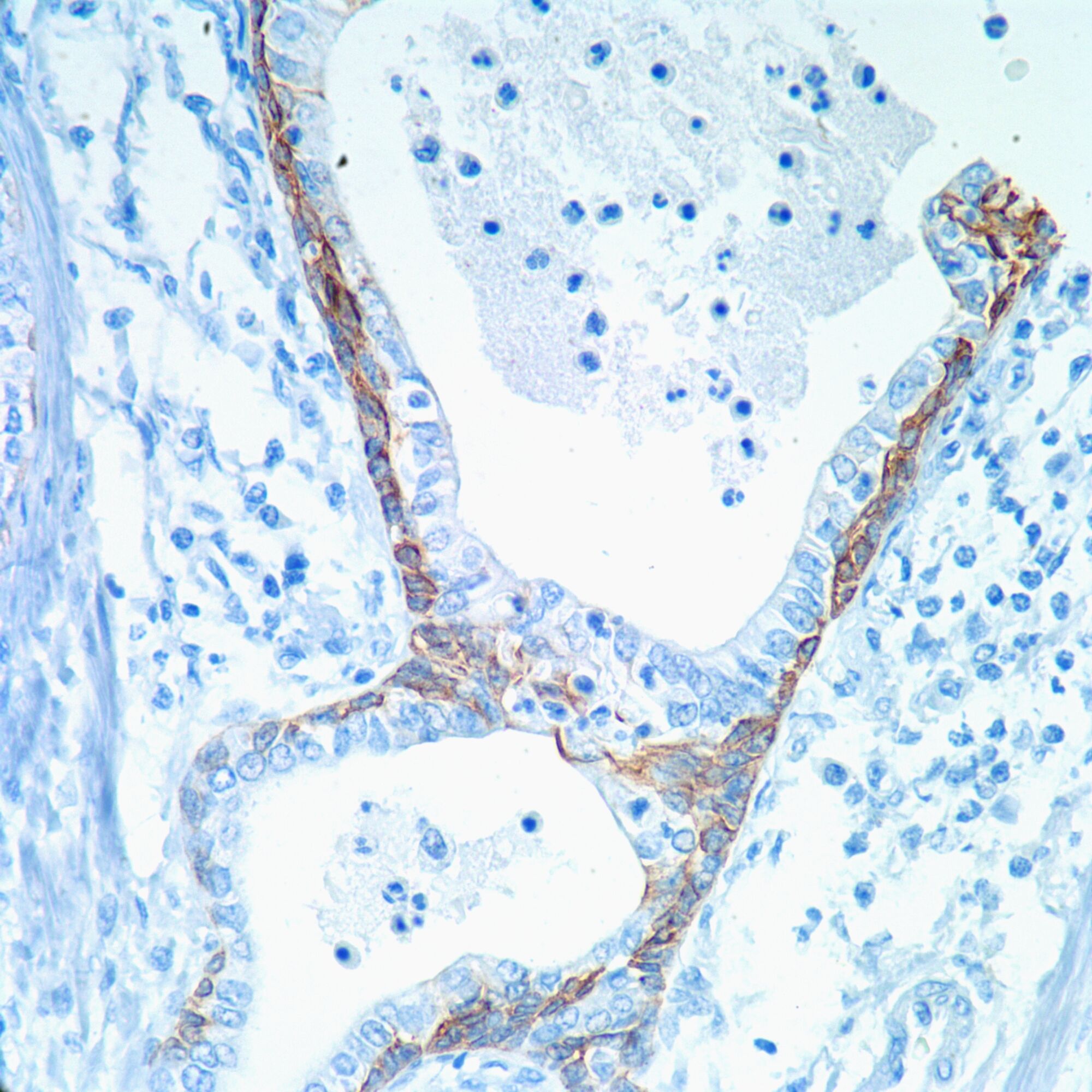


Cytokeratin 5 6 Ck 5 6 Pathology Resident Wiki Fandom
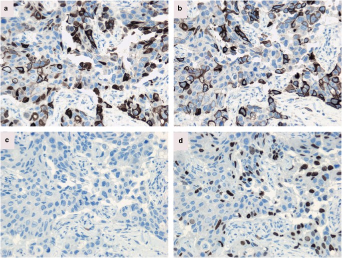


Cytokeratin 5 14 Positive Breast Cancer True Basal Phenotype Confined To Brca1 Tumors Modern Pathology


Breast Carcinoma Molecular Profiling And Updates Semantic Scholar


Ispub Com Ijpa 13 2


Prognostic Impact Of Egfr And Cytokeratin 5 6 Immunohistochemical Expression In Triple Negative Breast Cancer Sciencedirect


A Practical Approach To Undifferentiated Tumors A Review On Helpful Markers And Common Pitfalls Asian Archives Of Pathology
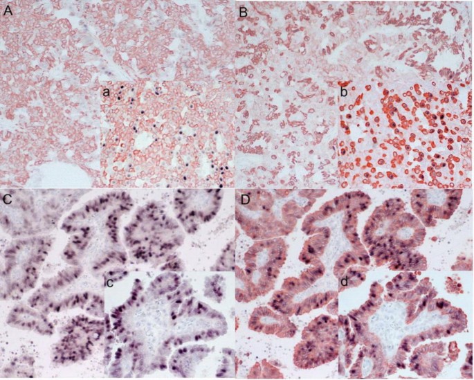


An Analysis Of Cyclin D1 Cytokeratin 5 6 And Cytokeratin 8 18 Expression In Breast Papillomas And Papillary Carcinomas Diagnostic Pathology Full Text


Immunohistochemistry Of Cytokeratin Ck 5 6 Cd44 And Ck As Prognostic Biomarkers Of Non Muscle Invasive Papillary Upper Tract Urothelial Carcinoma Jung 19 Histopathology Wiley Online Library


Best Practices Recommendations For Diagnostic Immunohistochemistry In Lung Cancer Journal Of Thoracic Oncology


Pathology Outlines Cytokeratin 5 6 And Ck5


Focal Positivity Of Immunohistochemical Markers For Pulmonary Squamous Cell Carcinoma In Primary Pulmonary Choriocarcinoma A Histopathological Study


Cytokeratin 5 6 P63 And Ttf 1 Immuno Marker Use In Tiny Non Small Cell Lung Cancer Medcrave Online


The Use Of P63 Immunohistochemistry For The Identification Of Squamous Cell Carcinoma Of The Lung


Utility Of 10 Immunohistochemical Markers Including Novel Markers Desmocollin 3 Glypican 3 S100a2 S100a7 And Sox 2 For Differential Diagnosis Of Squamous Cell Carcinoma From Adenocarcinoma Of The Lung Journal Of Thoracic Oncology


Expression Of Cytokeratin 5 6 In Epithelial Neoplasms An Immunohistochemical Study Of 509 Cases Modern Pathology


Cytogenetic Alterations And Cytokeratin Expression Patterns In Breast Cancer Integrating A New Model Of Breast Differentiation Into Cytogenetic Pathways Of Breast Carcinogenesis Laboratory Investigation
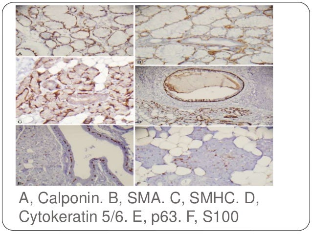


Review And Updates Of Immunohistochemistry In Selected Salivary Gland
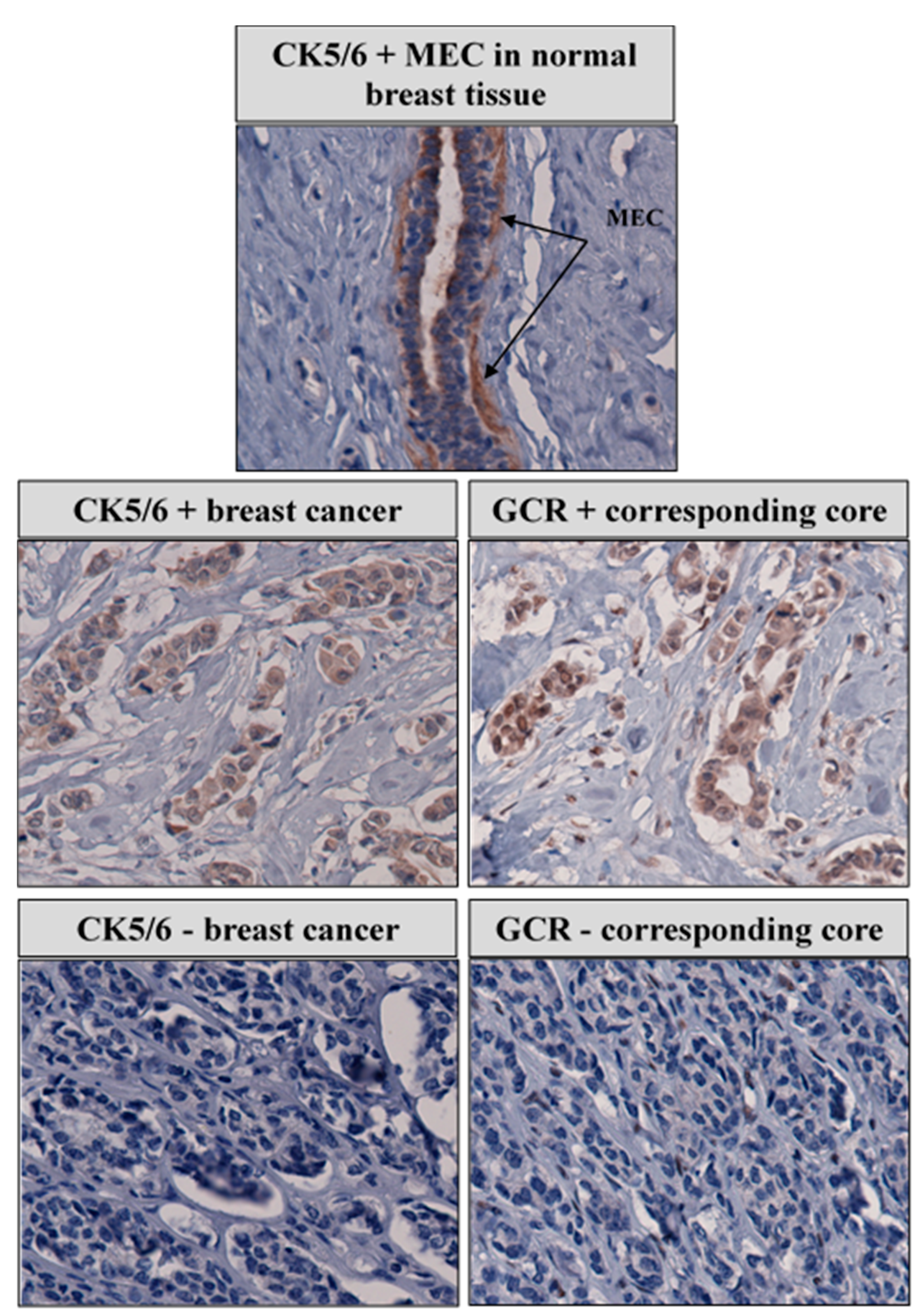


Cancers Free Full Text Genetic Variation And Immunohistochemical Localization Of The Glucocorticoid Receptor In Breast Cancer Cases From The Breast Cancer Care In Chicago Cohort Html
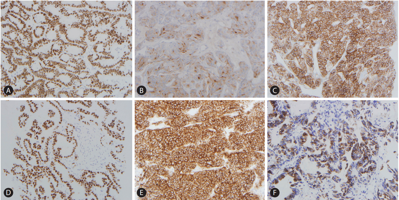


Pathologic Differential Diagnosis Of Metastatic Carcinoma In The Liver
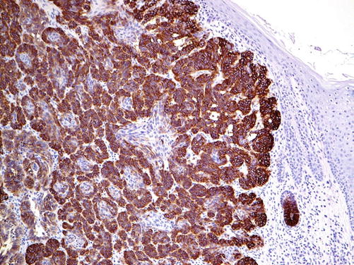


Markers And Immunoprofile Of Skin Tumors Springerlink


0 件のコメント:
コメントを投稿